Autonomous Matter
Quantitative Developmental Biology

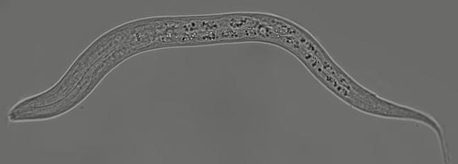
The Quantitative Developmental Biology group uses a quantitative, physics-inspired approach to study developmental biology. We work both on the small roundworm C. elegans, an important model organism for study of development, and in organoids, which are mouse or human mini-organs that can be grown in the lab.
Research focus
We are a highly interdisciplinary research group of physicists and biologists that study how, during animal development, robust patterns emerge in space and time, despite strong variability on the molecular and cellular level. Our approach is to study simple model systems for development by custom time-lapse microscopy, advanced AI-driven image analysis, quantitative data analysis, and predictive mathematical modeling.
In the nematode worm C. elegans, we study how cells correctly chose and maintain their cell type despite molecular variability and how the worm adjusts its developmental timing and progression in response to changing environmental conditions. In organoids, we study how individual cells divide, differentiate and rearrange to organize into a functional, self-maintaining tissue, with a focus on the mouse and human intestinal epithelium.
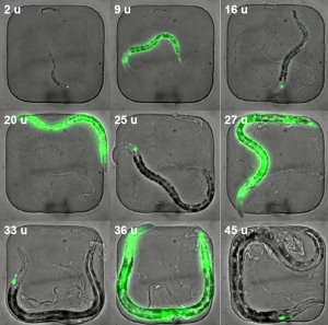
We have developed a microscopy approach based on a small microchamber to follow cell dynamics during the entire larval development of C. elegans worms, from hatching to adulthood, all while the animals themselves are growing, moving and feeding normally. We use this to study how cells control and adapt their behavior over time, as they first collectively build the animal’s body and subsequently maintain it, despite intrinsic variability at the molecular and cellular level and strong variations in external conditions, such as temperature and food availability. We focus on different aspects of these processes.
We study how bistable genetic switch networks maintain neuronal cell fate long after initial differentiation, without molecular fluctuations causing these networks to spontaneously de-differentiate. We also study how cells use molecular signals to record the passage of time and execute their dynamics at the right point in development. Using our ability to control the worm’s external environment, we investigate how the timing of cell dynamics adapts when adverse conditions slow development.
We discovered that when worms temporarily arrest larval growth and development due to external stresses, such as starvation, their cells execute transcription factor translocation pulses that are stochastic, yet highly synchronized throughout the body. We are studying how these pulses transmit stress information and maintain their synchronization.
Key publications:
- Long-term time-lapse microscopy of elegans post-embryonic development (Nature Communications, 2016)
- Mechanism of life-long maintenance of neuron identity despite molecular fluctuations (Elife, 2021)
- Temporal scaling in elegans larval development (PNAS, 2022)
- Timers, variability, and body-wide coordination: elegans as a model system for whole-animal developmental timing (Current Opinions in Genetics and Development, 2024)
- Body-wide synchronization of insulin-signaling dependent DAF-16/FOXO nuclear translocation pulses correlated with elegans growth (Nature Communications, 2025)
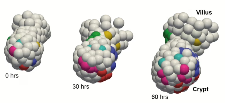
Even when we are adults, the stem cells in our organs keep on dividing and differentiating, to replenish cells that died – a process that is called homeostasis and is misregulated in many diseases. Organoid models now make it possible to visualize this process, that is normally hidden deep within the body, under the microscope. We use time-lapse microscopy to track the dynamics of all cells in organoids, as these cells divide, differentiate and die, with a focus on the intestinal epithelium.
Using such quantitative data, we study the developmental mechanisms that allow the intestine to maintain precise proportions and spatial patterns of stem cells and all differentiated cell types, even though the underlying processes, such a cell proliferation, differentiation and movements, show high intrinsic variability. We also study how mechanical interactions, due to cells pushing and pulling on each other as they divide and rearrange, controls the spatial patterning of differentiated cells and the removal of weak cells by extrusion from the epithelium.
Finally, we are increasingly turning to other organoid models, such as the mammary gland, and more complex organoid co-cultures to incorporate cells, such as fibroblasts and immune cells, that interact with the intestinal epithelium inside the body. All our work on organoids is done jointly with the group of Sander Tans, also at AMOLF.
Key publications:
- Mother cells control daughter cell proliferation in intestinal organoids to minimize proliferation fluctuations (eLife, 2022)
- Cell tracking for organoids: lessons from developmental biology (Frontiers in Cell and Developmental Biology, 2021)
- Organoid cell fate dynamics in space and time (Science Advances, 2023)
- Epithelial tension controls intestinal cell extrusion (Science, 2025)
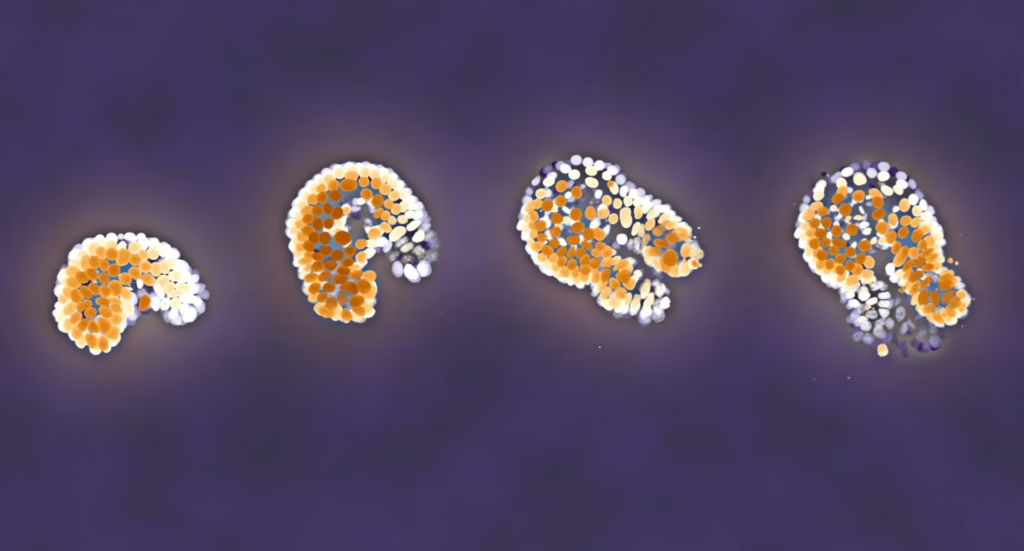
Key to our work is the tracking of cells in time, which for the number of cells in animals or organoids is simply impossible to do manually. We therefore put much effort in developing software for image analysis and cell tracking, leveraging the recent advances in AI and deep-learning to identify cells in microscopy images and track them through movements and divisions.
This has culminated in the recent development of OrganoidTracker 2.0, a cutting-edge cell tracker optimized for 3D organoid data, that uses advanced error rate prediction to enable users to only curate low-confidence track fragments, strongly reducing manual workload, or even perform fully automated analysis only of tracks that the algorithm deems high-confidence. We are constantly improving OrganoidTracker and are working to set up similar approaches for analyzing movies of C. elegans larval development.
Key publications:
- OrganoidTracker: Efficient cell tracking using machine learning and manual error correction (PLoS ONE, 2020)
- Label-free cell imaging and tracking in 3D organoids (Cell Reports Physical Sciences, 2025)
- Cell tracking with accurate error prediction (Nature Methods, 2025)





Quantitative Developmental Biology Latest news
Most recent publications