
Research highlights

Scientists discover how cells envelop bacteria

The protein GBP1 is a vital component of our body’s natural defence against pathogens. This substance fights against bacteria and parasites by enveloping them in a protein coat, but how the substance manages to do this has remained unknown until now. Researchers from Delft University of Technology and the AMOLF have now unravelled how this protein operates. This new knowledge, published in Nature Structural & Molecular Biology, could aid in the development of medications and therapies for individuals with weakened immune systems.

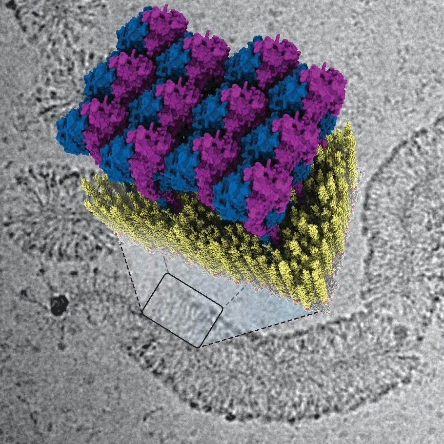
GBP1 proteins (blue and purple) attach to the membrane of a bacterium (yellow), zoomed in from an image taken with an electron microscope (in grayscale).

So-called Guanylate Binding Proteins (GBPs) play a crucial role in our innate immune system, explains biophysicist Arjen Jakobi: “GBPs form the first line of defence against various infectious diseases caused by bacteria and parasites. Examples of such diseases include dysentery, typhoid fever caused by Salmonella bacteria, and tuberculosis. The protein also plays a significant role in the sexually transmitted infection chlamydia as well as in toxoplasmosis, which is particularly dangerous during pregnancy and for unborn children.” 


Coat around bacteria
 

In their publication, Jakobi and his colleagues describe for the first time how the innate immune system fights against bacteria using GBP1 proteins. “The protein surrounds bacteria by forming a sort of coat around them,” explains Tanja Kuhm, PhD candidate in Jakobi’s research group and the lead author of the article. “By pulling this coat tighter, it breaks the membrane of the bacteria—the protective layer surrounding the intruder—after which immune cells can clear the infection.” 

Deciphering the defence strategy 

To decode the defence strategy of GBPs, the researchers examined how GBP1 proteins bind to bacterial membranes using a cryogenic electron microscope. This allowed them to see the process in great detail down to the scale of molecules. Jakobi: “We were able to obtain a detailed three-dimensional image of how the protein coat forms. Together with biophysical experiments conducted in Sander Tans’ research group at AMOLF, which enabled us to manipulate the system precisely, we succeeded in deciphering the mechanism of the antibacterial function.” 

By using an optical tweezer to hold a membrane vesicle under the microscope, PhD student Luca Gross at AMOLF was able to directly investigate the effect of GBP1. Gross: ‘In the experiments, I saw that the membrane vesicle was cut into small pieces after adding GBPI. It was fascinating to see how these proteins can autonomously exert force on the membrane to break it down’. 

Medications 

According to Jakobi, this research helps us understand better how our body is capable of combating bacterial infections. “If we can grasp this well, and we can specifically activate or deactivate the involved proteins through medication, it may offer opportunities to speed up getting rid of certain infections.” 

Reference

Tanja Kuhm, Clémence Taisne, Cecilia de Agrela Pinto, Luca Gross, Evdokia A. Giannopoulou, Stefan T. Huber, Els Pardon, Jan Steyaert, Sander J. Tans & Arjen J. Jakobi, Structural basis of antimicrobial membrane coat assembly by human GBP1, Nature Structural & Molecular Biology, 11 October (2024).

Contact

Do you have questions related to this research? Then please contact Sander Tans, email: S.Tans@amolf.nl.

 

Share article
What's happening

Most recent news items

All news items
Collaboration

Successful outcomes of international EBEAM program led by AMOLF

The EU Pathfinder project Electron Beams Enhancing Analytical Microscopy (EBEAM) that was recently completed has received a highly positive review of the European Innovation Council (EIC). EBEAM brought together eight European research institutions and companies, including AMOLF, that develop new concepts and instruments combining spectroscopic analysis with electron microscopy.

Read news item
Research highlights

Everything you need to know about organoids

Organoids are instrumental in improving our understanding of processes that are otherwise hidden inside the body. For instance, these small 3D organs can be used to test medicines on tissue grown from a patient’s own cells. AMOLF researchers carry out unique and complex experiments to follow organoids in time. In order to enable other researchers to benefit from their experiences, they now publish their methods in the prestigious journal Nature Protocols.

Read news item
Events and outreach

Minister Rianne Letschert wears hat and shoulder ornament inspired by Wim Noorduin’s research

This year at the opening of the parliamentary year (Prinsjesdag) Minister of Education, Culture and Science Rianne Letschert wears a spectacular hat and shoulder ornament. Both of them are inspired by the microscopic structures studied by group leader Wim Noorduin (AMOLF/UvA), which look remarkably like tiny flowers under a microscope. Artist and designer Malou Beemer translated these shapes, normally invisible to the naked eye, into wearable art.

Read news item
Nachi Stern, Group Leader Learning Machines at AMOLF
People and recognition

Nachi Stern awarded ERC Starting Grant to explore how matter learns

Why do brains learn, but rocks do not? Could a material one day adapt to its environment the way a living organism does? AMOLF group leader Dr. Nachi Stern has been awarded an ERC Starting Grant to investigate the physical laws of learning, in a project called, “Physical Learning in Dynamical Systems.”

Read news item
Stay informed

Get the latest research highlights, events, and news from our institute delivered to your inbox