Research highlights

Electron beam controls artificial optical molecule at the nanoscale

Metallic nanoparticles have the ability to confine light to volumes much smaller than the wavelength. If such particles are arranged in a special configuration they can show interesting optical phenomena, similar to a molecule consisting of multiple atoms. Such coupled particle systems are therefore referred to as metamolecules. Due to their small size, it is difficult to control and study their optical response. To address this problem, researchers from the FOM institute AMOLF and Stanford University have used an electron beam to control the optical behavior of such a metamolecule in a novel way by carefully positioning it with respect to the structure. The research was published in Nanoletters on October 12th, 2015.

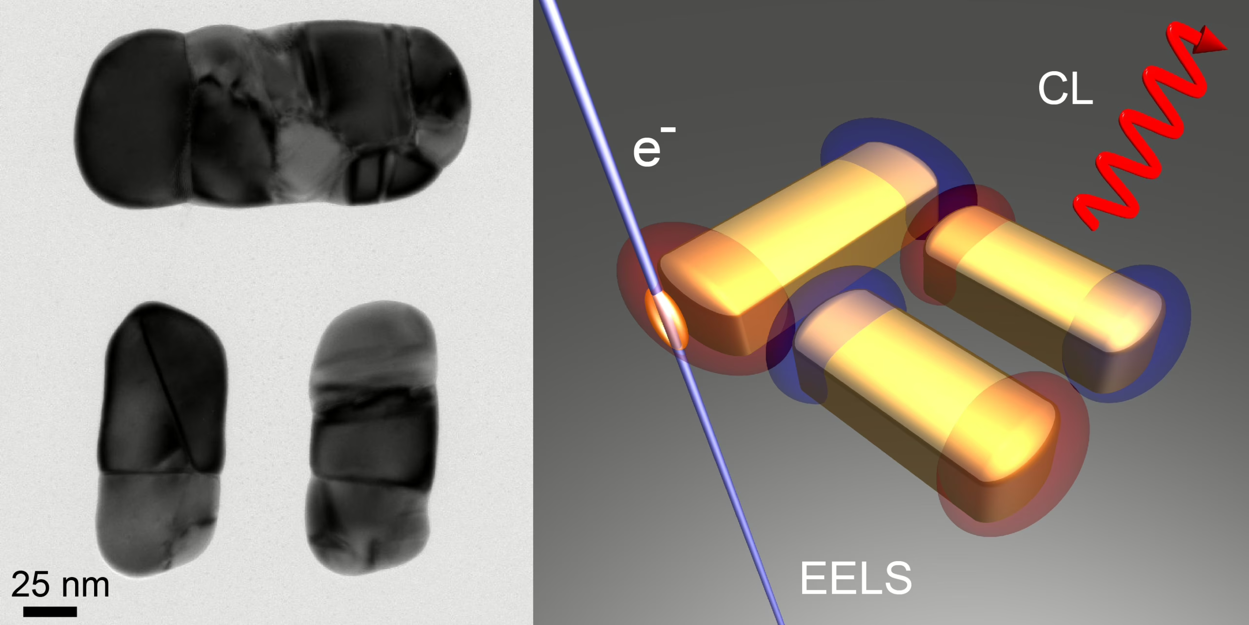
The study concerns a metamolecule consisting of one horizontal and two vertical gold nanoparticles, which is known as a dolmen structure (See Figure) due to its similarity in appearance with the ancient dolmen tombstones. This special arrangement of particles can render the structure transparent for certain colors whereas it strongly absorbs and scatters light for others, making it suitable to operate as a nanoscale optical sensing device or ruler.

The response of the structure depends on the intrinsic interaction between the particles which are dictated by the geometry, but also on how they are excited by an external stimulus. This stimulus can be a beam of light, but such a beam cannot be focused on a spot that is small enough, thus limiting the ability to precisely control the optical behavior of the dolmen.

As an alternative the researchers employed a tightly focused electron beam from an electron microscope to locally excite the dolmen. By carefully positioning the beam with respect to the structure, it is possible to selectively excite one or more elements in a controlled manner. Subsequently, they measured the resulting light emission (by using cathodoluminescence (CL) spectroscopy) and the energy loss experienced by the electron (by using electron energy loss spectroscopy (EELS)). By combining these two techniques the researchers were able to study what colors of light are efficiently emitted and which colors are absorbed in the dolmen when the structure is excited by the beam at different positions (see Figure for illustration). Controlling and studying the optical response in this way provides a powerful method which could be employed in fundamental nanophotonics research as well as optimization studies toward novel applications.

The research was carried out by Toon Coenen (AMOLF, now at Delmic BV), David Schoen, Sander Mann, Said Rodriguez, Benjamin Brenny, Albert Polman, and Mark Brongersma and was made possible by funding from the FOM Foundation, NanoNextNL, the Air Force Office of Scientific Research, and the European Research Council.

Reference:
T. Coenen, D.T. Schoen, S. A. Mann, S. R. K. Rodriguez, B. J. M. Brenny, A. Polman, and M. L. Brongersma
Nanoscale Spatial Coherent Control over the Modal Excitation of a Coupled Plasmonic Resonator System
Nano Lett. 15, (2015) | DOI: 10.1021/acs.nanolett.5b03614 (2015)

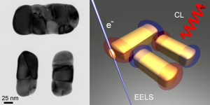
Figure: Left: Transmission electron microscopy image of the dolmen metamolecule. Right: Schematic illustration of the experiment. A tightly focused electron beam is used to locally excite the dolmen after which the light emission (CL) and the electron energy loss (EELS) are studied.
figure_Nanoletters Coenen
Share article
What's happening

Most recent news items

All news items
Collaboration

Successful outcomes of international EBEAM program led by AMOLF

The EU Pathfinder project Electron Beams Enhancing Analytical Microscopy (EBEAM) that was recently completed has received a highly positive review of the European Innovation Council (EIC). EBEAM brought together eight European research institutions and companies, including AMOLF, that develop new concepts and instruments combining spectroscopic analysis with electron microscopy.

Read news item
Research highlights

Everything you need to know about organoids

Organoids are instrumental in improving our understanding of processes that are otherwise hidden inside the body. For instance, these small 3D organs can be used to test medicines on tissue grown from a patient’s own cells. AMOLF researchers carry out unique and complex experiments to follow organoids in time. In order to enable other researchers to benefit from their experiences, they now publish their methods in the prestigious journal Nature Protocols.

Read news item
Events and outreach

Minister Rianne Letschert wears hat and shoulder ornament inspired by Wim Noorduin’s research

This year at the opening of the parliamentary year (Prinsjesdag) Minister of Education, Culture and Science Rianne Letschert wears a spectacular hat and shoulder ornament. Both of them are inspired by the microscopic structures studied by group leader Wim Noorduin (AMOLF/UvA), which look remarkably like tiny flowers under a microscope. Artist and designer Malou Beemer translated these shapes, normally invisible to the naked eye, into wearable art.

Read news item
Nachi Stern, Group Leader Learning Machines at AMOLF
People and recognition

Nachi Stern awarded ERC Starting Grant to explore how matter learns

Why do brains learn, but rocks do not? Could a material one day adapt to its environment the way a living organism does? AMOLF group leader Dr. Nachi Stern has been awarded an ERC Starting Grant to investigate the physical laws of learning, in a project called, “Physical Learning in Dynamical Systems.”

Read news item
Stay informed

Get the latest research highlights, events, and news from our institute delivered to your inbox