A team of AMOLF, Thermo Fisher Scientific and Delmic has constructed two new microscopes that can take optical images at the nanoscale, with a time resolution down to 1 ps. The three partners formed a consortium in 2016, with the aim to integrate a Scanning Electron Microscope (SEM) and a light collection and analysis system into a new microscope in which the electron beam is pulsed and the generated light (cathodoluminescence) is collected in a time-resolved way. During the final project meeting with the three partners on May 22, 2018, the two new ultrafast SEMs were presented. One of the two new instruments is already brought on the market by Thermo Fisher and Delmic; the first instrument has been sold in April 2018. The new ultrafast SEMs will be presented at the International Microscopy Conference in Sydney (September 9-14, 2018).
Ultrafast beam-blanker
The first ultrafast SEM is composed of a Thermo Scientific Quanta 650 FEG SEM and a Delmic SPARC light collection and analysis system, with an ultrafast electrostatic beam blanker integrated in the SEM electron column. Using dedicated electronics and software the system can deliver electron pulses shorter than 30 ps (at 5 keV), while the time dependence of the cathodoluminescence is collected using single-photon counting/correlation spectroscopy. The new instrument delivers two-dimensional images of the cathodoluminescence lifetime and g(2) photon correlations. Such data provide key information for the study of light emission characteristics of semiconductor nanostructures and quantum optical studies in a broad range of systems.
Pulsed-laser driven cathode
In the second microscope the SEM electron cathode is excited by 250-femtosecond UV laser pulses, creating ultrashort electron pulses. This enables spatial imaging of optical phenomena at picosecond time scales. In addition, this microscope enables ultrafast pump-probe spectroscopy, in which the laser pulse is split in two parts: one excites a sample, while the other excites the photocathode, generating an electron pulse that probes the sample. The combination of ultrafast pump-probe cathodoluminescence spectroscopy with the very high spatial resolution makes this a unique instrument.
The collaboration has led to two published papers:
Nanoscale relative emission efficiency mapping using CL g(2) imaging
S. Meuret, T. Coenen, S. Woo, Y.-H. Ra, Z. Mi and A. Polman, Nano Lett. 18, 2288 (2018)
Photon bunching reveals single-electron cathodoluminescence excitation efficiency in InGaN quantum wells
S. Meuret, T. Coenen, M. Lätzel, S. Christiansen, S. Conesa Boj, and A. Polman, Phys. Rev. B 96, 035308 (2017)
A third paper that describes the technical aspects of the new microscopes has been submitted:
Complementary cathodoluminescence lifetime imaging configurations in scanning electron microscopy
S. Meuret, T. Coenen, M. Solà-Garcia, E. Kieft, H. Zeijlemaker, M.Latzel, S. Christiansen, S.Y. Woo, Y-H Ra, Z. Mi, A. Polman.
For details on cathodoluminescence microscopy research in the group of Albert Polman at AMOLF, see: http://www.erbium.nl/arcis.
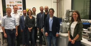
AMOLF, Thermo Fisher and Delmic project team at the new ultrafast SEM. From left to right: Erik Kieft (Thermo Fisher), Ernst Jan Vesseur (Thermo Fisher), Nico Clemens (Thermo Fisher), Sophie Meuret (AMOLF), Toon Coenen (Delmic/AMOLF), Albert Polman (AMOLF), Sander den Hoedt (Delmic), Andries Effting (Delmic) and Magda Sola Garcia (AMOLF).