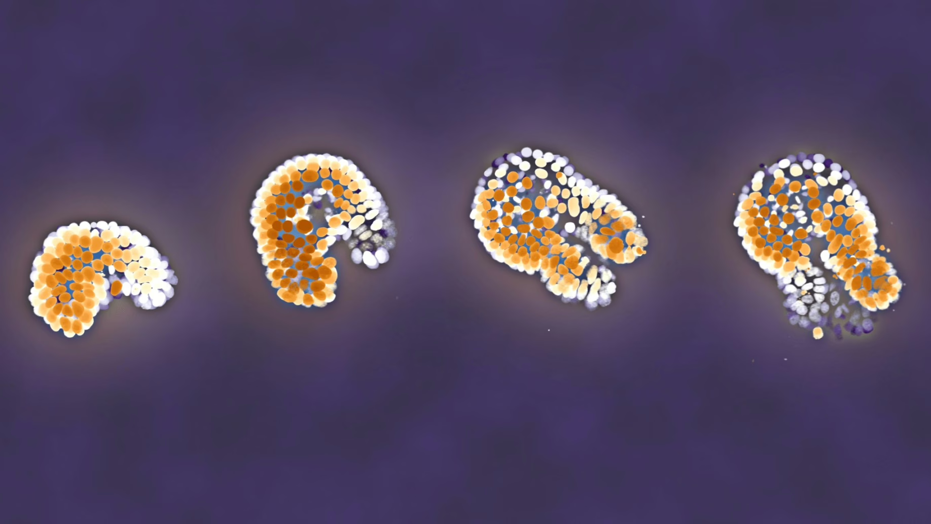
Left: Microscope images of cell nuclei. The cell nuclei have been made fluorescent by means of genetic modification. They have been digitally colored based on their depth.
Right: Digital reconstruction of the cells followed (see video left). Each sphere represents a single cell. Spheres with the same color descend from the same ancestral cell. From this, we can see that the blue cell has divided into 12 descendants at the end of the video.
Intestinal cells are among the fastest growing and dividing cells in the body. But if all intestinal cells were to divide and their descendants too, then the result would be exponential growth that can cause tumors or diseases. That does not happen; cell growth in the intestines is constant and fluctuates little. The intestines must therefore have a mechanism to regulate that growth.
To investigate how that comes about, Jeroen van Zon, group leader Quantitative Developmental Biology, Sander Tans (Biophysics) and Jacco van Rheenen from the Netherlands Cancer Institute and Oncode Institute developed a model system of an intestine, the so-called organoid. This is a cultured but entirely functioning piece of mini intestine. Just like in a real intestine there are cavities (crypts) in which stem cells are located that divide and differentiate into, for example, intestinal tissue.
Using a microscope, the researchers could examine each individual cell in the mini intestine and take a photo every ten minutes. That had not previously been done. This approach allowed them to follow which cells divided, and from this, they derived a lineage. The researchers’ first question was whether such an organoid was indeed a good model for cell growth. Is the cell growth in an organoid just as constant as in a real intestine? Van Zon: “I was surprised to discover that this is indeed the case. Clearly, the organoid does not need the rest of the body to achieve stable cell growth.”
Preference for symmetry
The researchers could also establish which cells divided: both daughters of a mother cell, one of the two or neither. If both daughters do the same, then that is called symmetric proliferation (growth); if one divides and the other does not, then that is called asymmetric proliferation. The cells in the mini intestine were found to have a strong preference for symmetric growth. “That is at odds with the classical model, which states that completely asymmetric growth gives rise to the least fluctuations,” says Van Zon. “It looks like the opposite happens in a realistic model of the intestine: symmetric proliferation gives rise to the least fluctuations.”
The researchers also demonstrated that an organoid is a good model for a real intestine via collaboration with the Oncode institute and the Netherlands Cancer Institute. In living mice, they can count and follow intestinal cells, albeit in less detail than in the organoids. If, after a certain period, a cell has an equal number of descendants, then that is an indication of symmetric growth. And that is precisely what the researchers found. “This once again shows that the organoid is a good model system for the real intestine,” states Van Zon.
Why do twins do the same?
Finally, the question arises as to why two sister cells from the same mother cell behave in the same way. In the language of Twin Studies: is that a matter of nature or nurture? Do the daughters receive a signal from the mother as a result of which they either both divide or not? Or is the behavior determined by the fact that they both grow in the same environment, where, for example, they are exposed to signaling molecules? Paneth cells in the intestine are known to give off such signals.
To find out, the researchers followed all cells not just temporally, but also spatially. That allowed them to see whether sister cells always stayed at exactly the same location. For example, one of them was sometimes located a bit further from the Paneth cells. Yet despite that distance, the two sister cells nevertheless did the same, namely, they exhibited symmetric behavior. “From this, we conclude that a signal they receive from the mother cell determines whether the daughter cells divide or not,” says van Zon. “I think that is one of the most unusual outcomes from this research: that the division of cells in the intestines is strictly symmetrical because of the signal given by their mother cell.”
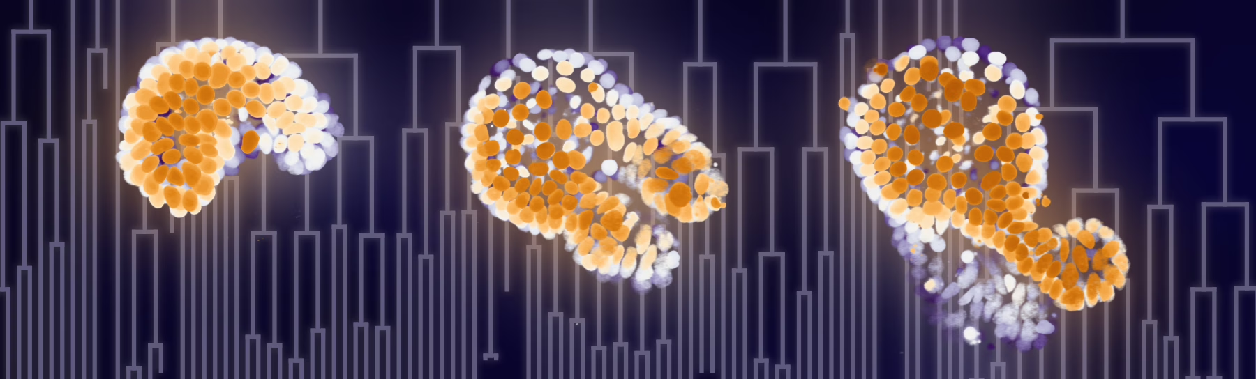
Fluorescence microscopy images of the cell nuclei of a growing organoid. The organoid has protrusions where stem cells are found: the crypts. Initially, the inside of the organoid is almost empty and contains just water. Over the course of time, it becomes filled with old, dead cells. The cell nuclei have been made fluorescent by means of genetic modification.
Reference
Guizela Huelsz-Prince, Rutger N.U. Kok, Yvonne J. Goos, Lotte Bruens, Xuan Zheng, Saskia I. Ellenbroek, Jacco van Rheenen, Sander J. Tans , Jeroen S. van Zon, Mother cells control daughter cell proliferation in intestinal organoids 1 to minimize proliferation fluctuations, eLife, November 30 (2022).
doi.org/10.7554/eLife.80682