The Quantitative Developmental Biology group uses the nematode C. elegans as a model system to gain a better understanding of developmental processes. Van Zon explains: ‘The adult nematodes are composed of about 1000 cells and many of their basic mechanisms are very similar to those of more complex animals and humans. Furthermore, the nematodes are transparent, which makes them highly suitable for studying under a microscope.’
Variation
Van Zon and Huelsz-Prince studied a group of five cells that develop into the female reproductive organ of the – usually hermaphroditic – animal during the nematode’s “puberty”. ‘The five cells that develop into the vulva communicate with the anchor cell in order to choose one of three possible cell types – primary, secondary, tertiary – depending on the distance to the anchor cell,’ explains Van Zon. ‘Contrary to what we expected, we saw a large variation in the position of the anchor cell with respect to the cells, whereas the final pattern that they assumed was always the same.’
Canalisation
An important idea in developmental biology is that this arises due to canalisation: a sort of feedback mechanism in the cells, which ensures that the development of organisms is robust and reproducible. Exactly how that feedback mechanism works used to be a mystery. ‘But, now, we have seen canalisation actually taking place for the first time,’ explains Van Zon. ‘If an anchor cell is in the “wrong” position, then in theory, two cells have an almost equal chance of choosing the primary cell type. In such a case, we saw the cells shifting so that a single cell once again came to lie closest to the anchor cell. Furthermore, the cells “communicate” with each other: the cell that chooses the primary cell type under the influence of the anchor cell transmits signal molecules to its neighbours, as a result of which they do not choose the primary cell type. By making these molecules fluorescent, we could observe the communication between the cells under a microscope. As a result of the experiments and the mathematical models that we used to analyse these, we now have a better understanding of how the combination of signals that the cells transmit to each other and the migration to the correct position, form the feedback mechanism for canalisation.’
The next step
One remaining unanswered question is the order in which the different processes take place. For the research in this publication, Van Zon and Huelsz-Prince used dead nematodes that were observed in various stages of development. The group has since developed a technique that allows them to study living nematodes in a comparable manner, so that they can follow the animal’s development over time. Van Zon: ‘With this technique, we can look even more closely at the exact timing of the processes that give rise to canalisation. We want to use this new technique to unravel canalisation in other developmental processes as well.’
Reference
Guizela Huelsz-Prince and Jeroen Sebastiaan van Zon, Canalization of C. elegans Vulva Induction against Anatomical Variability, Cell Systems 4, 219–230 (2017) | DOI: 10.1016/j.cels.2017.01.009
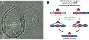
Fig. caption:
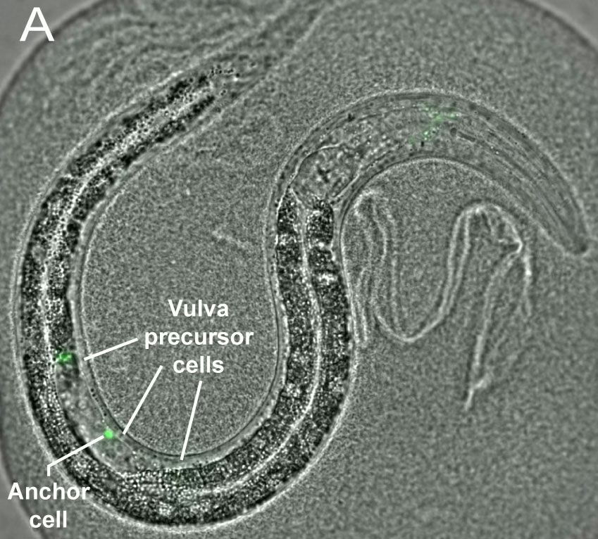
A) A C.elegans worm with the anchor cell (marked green with fluorescent proteins) and the three closest vulva cells.
B) Researchers found a large variation in the position of the anchor cell ( purple) relative to the vulva cells in the development of C. elegans. On the left, the cell nearest to the anchor cell (red violet) receives the strongest signal to acquire the primary fate. It subsequently sends signals to its neighbours (the so-called Notch signal), which ensures that these develop into secondary cells (blue).
On the right, two cells have a virtually equal chance of becoming the primary cell type. The cell that is slightly closer creeps towards the anchor cell and sends a slightly stronger Notch signal to the neighbouring cell. The combination of these two effects ensures canalisation: despite the initial variation, the final pattern is the same as that in the left-hand part of the figure.