Research highlights

How do hydrophobins work at the water surface?

Hydrophobins are a group of highly surface-active proteins that are produced by fungi. They are known for their unique functions related to interfaces. Hydrophobins largely reduce the surface tension of water, strongly adhere to surfaces and form protective surface coatings, all functions that play important roles in fungal physiology. Hydrophobins are used in several industrial applications, such as foams, dispersions and functional coatings. These applications rely on the unique surface properties of hydrophobins characterized by the proteins’ self-assembly into robust films.

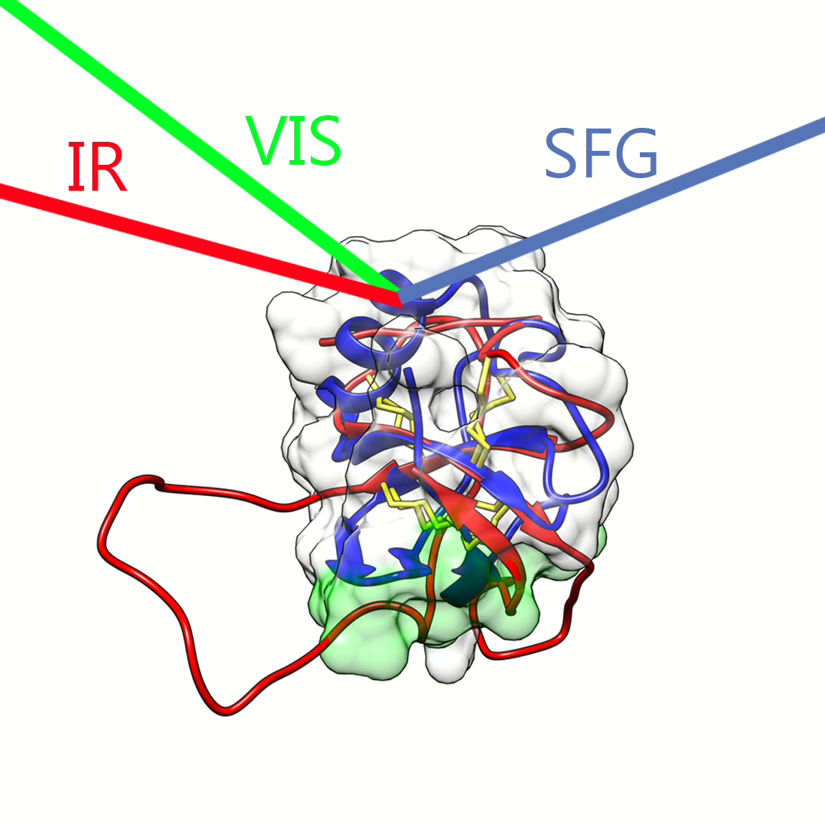
Schematic of hydrophobin protein studied with surface-specific sum-frequency generation (SFG) spectroscopy.

New discoveries
While much progress has been made in the characterization of the macroscopic structure of hydrophobin films, little was known about the initial stages of film formation and the microscopic structure of these extraordinary proteins directly at the water surface. Even less information was available on their dynamics, such as conformational fluctuations or intermolecular and solvent interactions.  Recently, researchers from the group of Huib Bakker  and collaborators at the VTT Technical Research Centre in Finland were able to uncover some of these mysteries. They investigated the mechanisms using the advanced (nonlinear) technique of surface-specific sum-frequency generation (SFG) spectroscopy. This technique is used to investigate the protein properties through their intrinsic molecular vibrations. Using SFG spectroscopy the researchers were able to prove that the two mayor hydrophobin classes have different self-assembling mechanisms at the interface. The findings were published online on September 30th in the Journal of Physical Chemistry Letters.

Functional Amyloids
For class 1 hydrophobins a surface-driven self-assembly mechanism was found in which the central β-barrel structure remains intact and stacks into a larger scale architecture, amyloid-like rodlets. These findings are significant, because class 1 hydrophobins are one of the few known examples in nature were amyloid-like structures have a positive function and are not associated with a disease. For class 2 hydrophobins no structural changes were observed, which agrees with the picture that class 2 hydrophobins do not form amyloid-like rodlets, but instead form films with a hexagonal structure and distinct repeating units.

Reference
Konrad Meister, Alexander Bäumer, Geza R. Szilvay, Arja Paananen and Huib J. Bakker,  Self-Assembly and Conformational Changes of Hydrophobin Classes at the Air–Water Interface,  The Journal of Physical Chemistry Letters, doi: 10.1021/acs.jpclett.6b01917

Share article
What's happening

Most recent news items

All news items
Collaboration

Successful outcomes of international EBEAM program led by AMOLF

The EU Pathfinder project Electron Beams Enhancing Analytical Microscopy (EBEAM) that was recently completed has received a highly positive review of the European Innovation Council (EIC). EBEAM brought together eight European research institutions and companies, including AMOLF, that develop new concepts and instruments combining spectroscopic analysis with electron microscopy.

Read news item
Research highlights

Everything you need to know about organoids

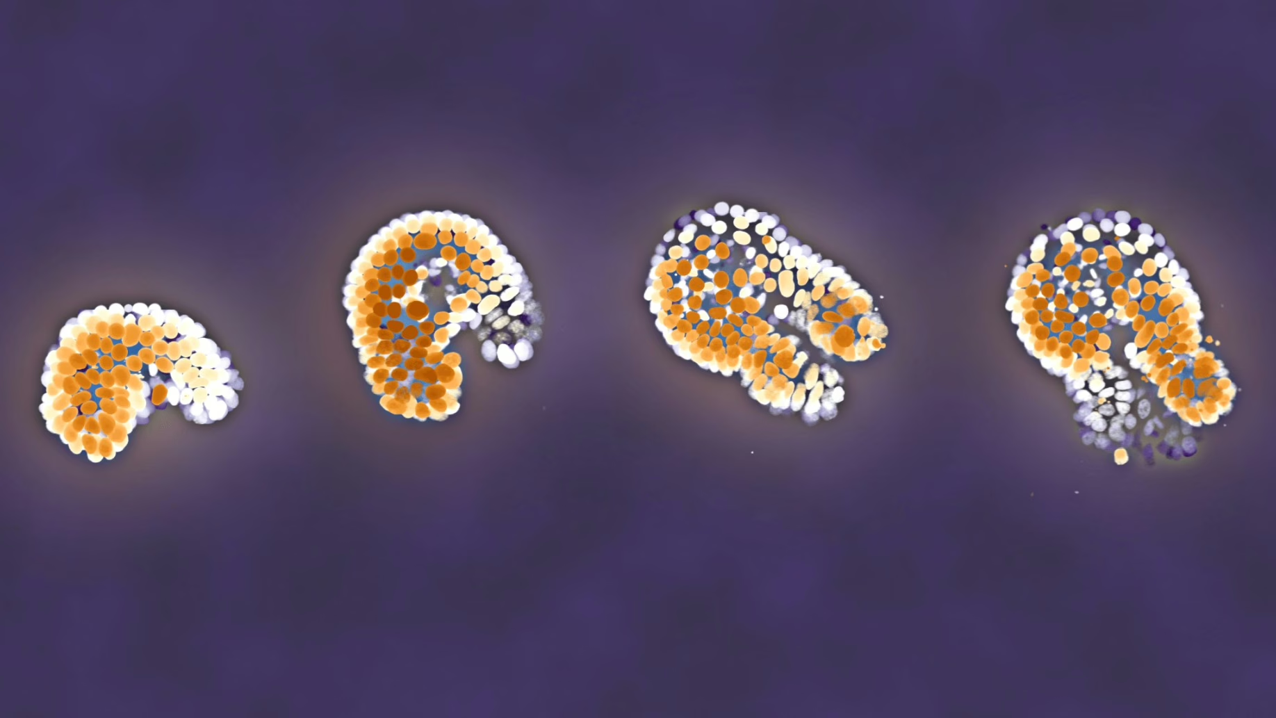
Organoids are instrumental in improving our understanding of processes that are otherwise hidden inside the body. For instance, these small 3D organs can be used to test medicines on tissue grown from a patient’s own cells. AMOLF researchers carry out unique and complex experiments to follow organoids in time. In order to enable other researchers to benefit from their experiences, they now publish their methods in the prestigious journal Nature Protocols.

Read news item
Events and outreach

Minister Rianne Letschert wears hat and shoulder ornament inspired by Wim Noorduin’s research

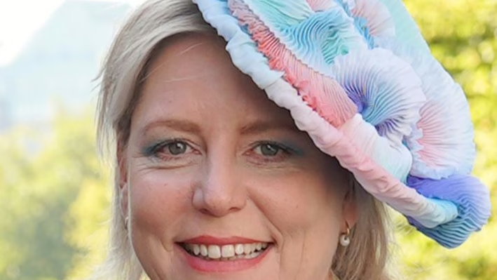
This year at the opening of the parliamentary year (Prinsjesdag) Minister of Education, Culture and Science Rianne Letschert wears a spectacular hat and shoulder ornament. Both of them are inspired by the microscopic structures studied by group leader Wim Noorduin (AMOLF/UvA), which look remarkably like tiny flowers under a microscope. Artist and designer Malou Beemer translated these shapes, normally invisible to the naked eye, into wearable art.

Read news item
Nachi Stern, Group Leader Learning Machines at AMOLF
People and recognition

Nachi Stern awarded ERC Starting Grant to explore how matter learns

Why do brains learn, but rocks do not? Could a material one day adapt to its environment the way a living organism does? AMOLF group leader Dr. Nachi Stern has been awarded an ERC Starting Grant to investigate the physical laws of learning, in a project called, “Physical Learning in Dynamical Systems.”

Read news item
Stay informed

Get the latest research highlights, events, and news from our institute delivered to your inbox