Research highlights

Nanoscale optical tomography using electrons

A team of researchers from AMOLF and Stanford University has developed a novel way to make three-dimensional optical images of nanoscale objects. They irradiate the object with a 30 keV electron beam under different angles and collect the cathodoluminescence emitted by the sample. Using a tomographic reconstruction they then determine the three-dimensional light distribution in metallic crescent particles at nanoscale resolution.

Cathodoluminescence
Many novel optical devices, such as solar cells, LEDs or lasers require control over the concentration of light at the nanoscale. However, standard optical microscopy cannot be used to probe light at this scale as the imaging resolution is limited by the optical diffraction limit (~250 nanometer). A new instrument, developed in Albert Polman’s group at AMOLF in the past years, uses an electron beam to excite materials and detects the emitted light (called cathodoluminescence) to make optical images at a resolution far below the diffraction limit. So far, however, only two-dimensional images were made.

Tomography
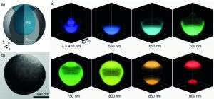
The new study, published in Nature Nanotechnology on April 6th, 2015, expands this technique to carry out three-dimensional tomography. Combining expertise from the groups at AMOLF and Stanford, the team uses 30 keV electrons to excite crescent-shaped core-shell particles composed of a polystyrene core and a gold shell. These particles, with a diameter of 250 nanometer, serve as a model system in which multicolored light is concentrated in different nanoscale focal spots. The research team irradiated crescents with electrons under multiple angles, collected the emitted light and carried out a tomographic reconstruction. In this way multicolored optical hot spots were imaged in three dimensions at a resolution smaller than the diffraction limit.

The Stanford part of the team was led by Jennifer Dionne, an assistant professor in Stanford’s Department of Materials Science and Engineering. Dionne is a long-term collaborator of AMOLF in the field of plasmonics and optical metamaterials. Dionne and Polman: “This was an exceptional project that became successful because of the complementary expertise of all of us. Together we are exploring new application areas that are opening up. We are already applying the images in the characterization of solar cells. We also think our new technique could be modified for imaging biological systems without the need for fluorescent labels.”

Reference
A. Atre, B.J.M. Brenny, T. Coenen, A. Polman and J.A. Dionne, Nanoscale optical tomography with cathodoluminescence spectroscopy Nature Nanotech. 10, 429–436 (2015) | DOI: 10.1038/nnano.2015.39
figuur a b c CL tomography crescent4

3D tomograms of nanoscale light (a) Schematic of crescent particle composed of a polystyrene core and Au shell. (b) Transmission electron micrograph of a crescent particle, showing a sharp Au edge revealing the bare polystyrene core at the top-right corner. (c) Cathodoluminescence tomograms at different wavelengths. A blue optical hot spot is observed at the bottom center inside the particle while a red hotspot is observed within the cusp at the top.
2015_credits AMOLF_Stanford_Tremani_lowres

© AMOLF/Stanford/Tremani

Share article
What's happening

Most recent news items

All news items
Collaboration

Successful outcomes of international EBEAM program led by AMOLF

The EU Pathfinder project Electron Beams Enhancing Analytical Microscopy (EBEAM) that was recently completed has received a highly positive review of the European Innovation Council (EIC). EBEAM brought together eight European research institutions and companies, including AMOLF, that develop new concepts and instruments combining spectroscopic analysis with electron microscopy.

Read news item
Research highlights

Everything you need to know about organoids

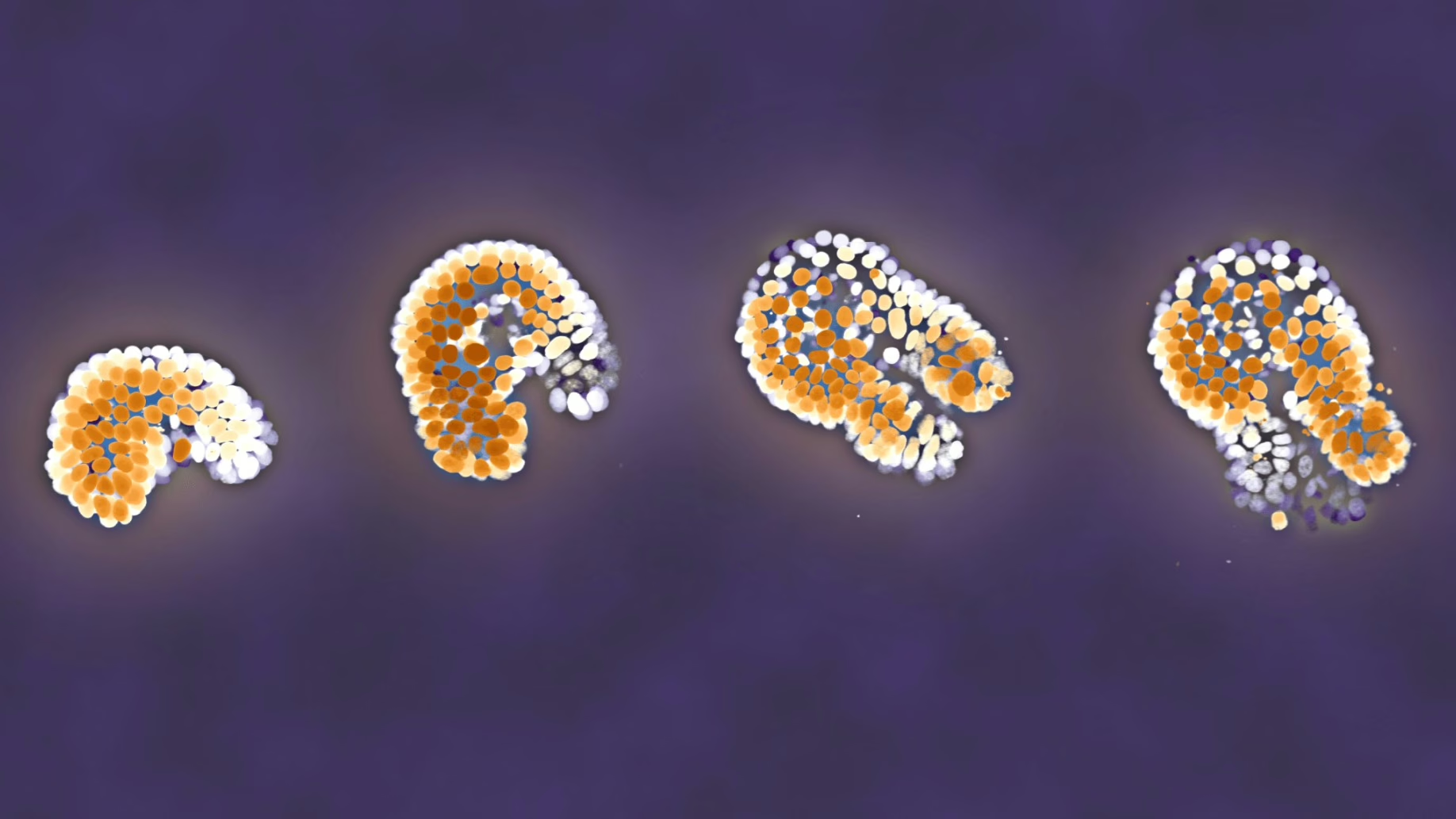
Organoids are instrumental in improving our understanding of processes that are otherwise hidden inside the body. For instance, these small 3D organs can be used to test medicines on tissue grown from a patient’s own cells. AMOLF researchers carry out unique and complex experiments to follow organoids in time. In order to enable other researchers to benefit from their experiences, they now publish their methods in the prestigious journal Nature Protocols.

Read news item
Events and outreach

Minister Rianne Letschert wears hat and shoulder ornament inspired by Wim Noorduin’s research

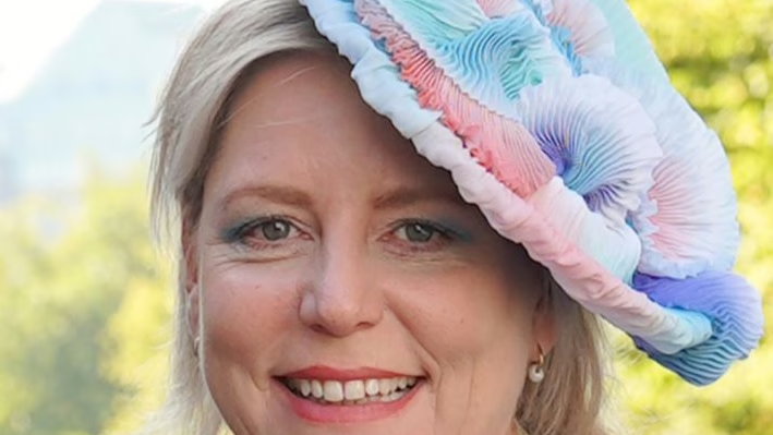
This year at the opening of the parliamentary year (Prinsjesdag) Minister of Education, Culture and Science Rianne Letschert wears a spectacular hat and shoulder ornament. Both of them are inspired by the microscopic structures studied by group leader Wim Noorduin (AMOLF/UvA), which look remarkably like tiny flowers under a microscope. Artist and designer Malou Beemer translated these shapes, normally invisible to the naked eye, into wearable art.

Read news item
Nachi Stern, Group Leader Learning Machines at AMOLF
People and recognition

Nachi Stern awarded ERC Starting Grant to explore how matter learns

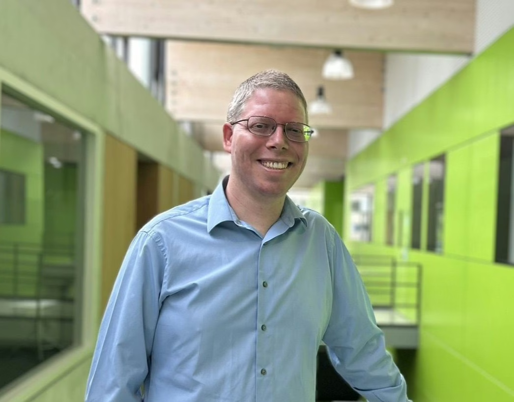
Why do brains learn, but rocks do not? Could a material one day adapt to its environment the way a living organism does? AMOLF group leader Dr. Nachi Stern has been awarded an ERC Starting Grant to investigate the physical laws of learning, in a project called, “Physical Learning in Dynamical Systems.”

Read news item
Stay informed

Get the latest research highlights, events, and news from our institute delivered to your inbox