Information in Matter
Biophysics

The Biophysics group develops novel biophysics methods to study dynamical processes in biology. We focus specifically on how proteins are made by ribosomes, and how cells work together to form organs. With this approach, we aim to understand the underlying mechanisms of cellular failure and disease.
Research focus
Researchers in the Biophysics research group are fascinated by the remarkable dynamic organization of living systems. We focus specifically on the cellular dynamics critical to organs, using organoids (mini lab-grown organs) and AI-driven imaging, and on the dynamics of ribosomes and chaperones, responsible for building and folding of proteins. To study cellular dynamics, we use single-molecule and sequencing methods.
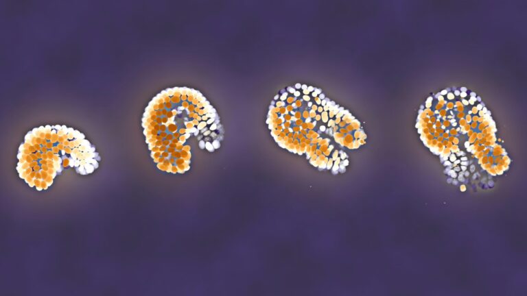
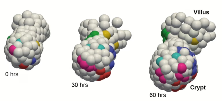
Organoids are assemblies of cells that recapitulate organ shape and function. Organoids are currently revolutionizing many aspects of cellular biology, as they allow for the study of disease-relevant processes outside the body. However, technical challenges have long prevented cellular dynamics from being quantified from time-lapse movies. Over the past years, we have developed AI-driven analysis methods to track all cells within organoids as they grow, divide, move, differentiate into different functional cell types, and ultimately die.
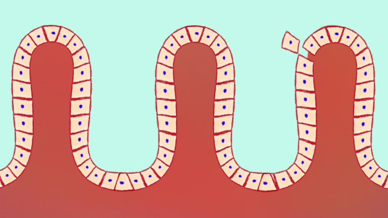
This tool shows the truly fascinating ways in which all these systems self-organize into functional tissues. For instance, we have shown that highly dynamic cross-cellular actin structures detect mechanically weak cells to extrude them from the intestinal epithelium, and that cellular identity choices are not governed by a spatial gradient of signaling molecules but rather by a timer mechanism that starts ticking when two key cell types dissociate from each other. This approach can be broadly applied to different organ cell types, organ systems, but also immune cells, which we are also exploring. We do our organoid work together with AMOLF colleague Jeroen van Zon.
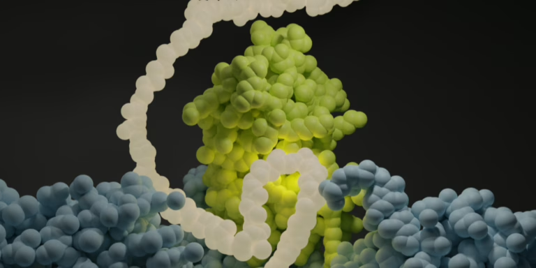
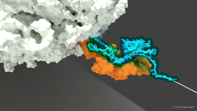
Polysomes are RNA messages decorated by ribosomes, as well as the proteins they produce and other cofactors. While known for decades, their functional purpose has long remained unclear. Recent work by us and others now suggest that polysomes enable cooperation between ribosomes. We mainly use single-molecule tools to mechanistically understand this cooperation. We also exploit RNA sequencing methods to characterize it across the proteome in cells, in collaboration with the Bukau-Kramer lab in Heidelberg.
For instance, we have shown how two ribosomes are physically linked via the proteins they are synthesizing, and hence jointly produce protein dimers. Strikingly, such nascent chain interactions can suppress misfolding. These and other findings indicate a wealth of dynamics and regulation occurring within polysomes, which we are exploring. These processes may be relevant to all major cellular processes, as well as to mRNA vaccines and AI-generated protein design.
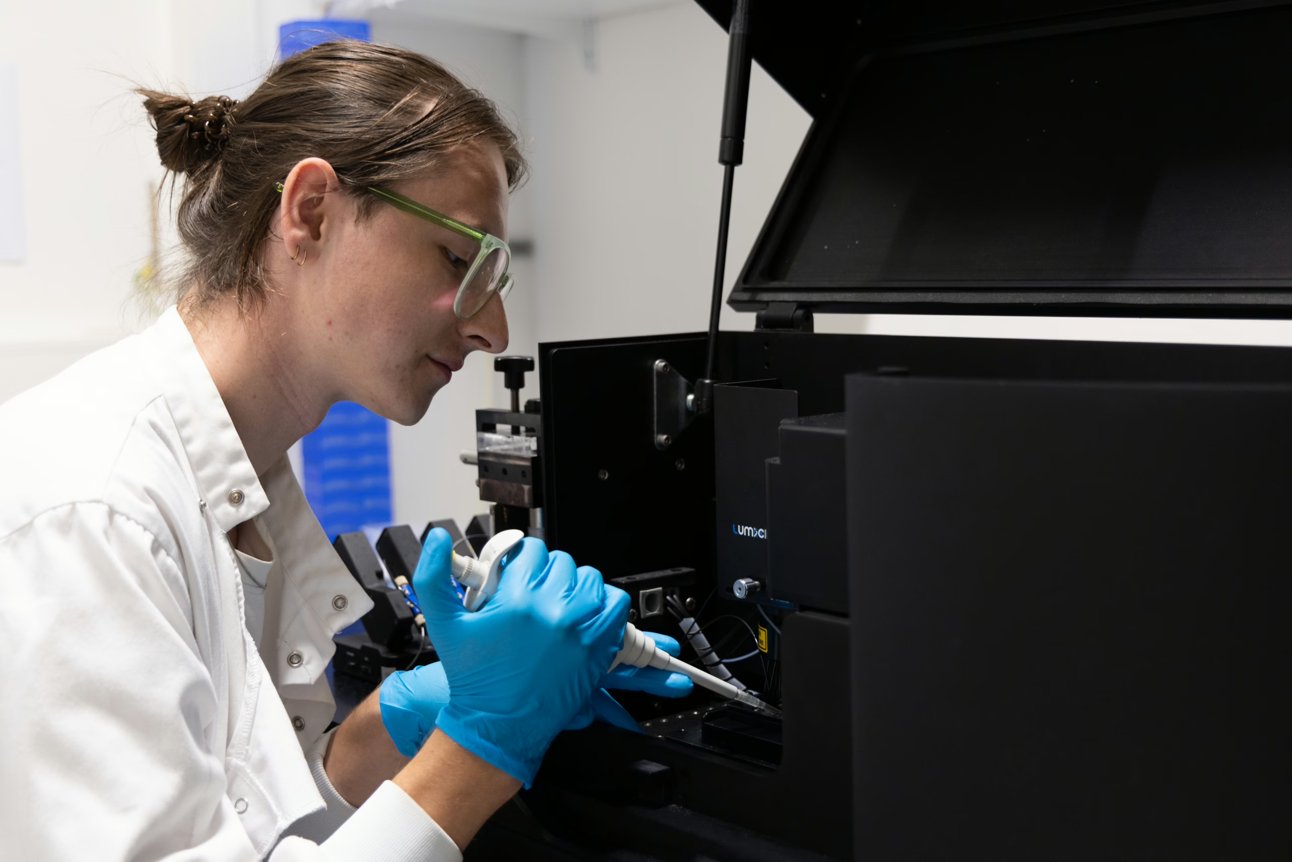
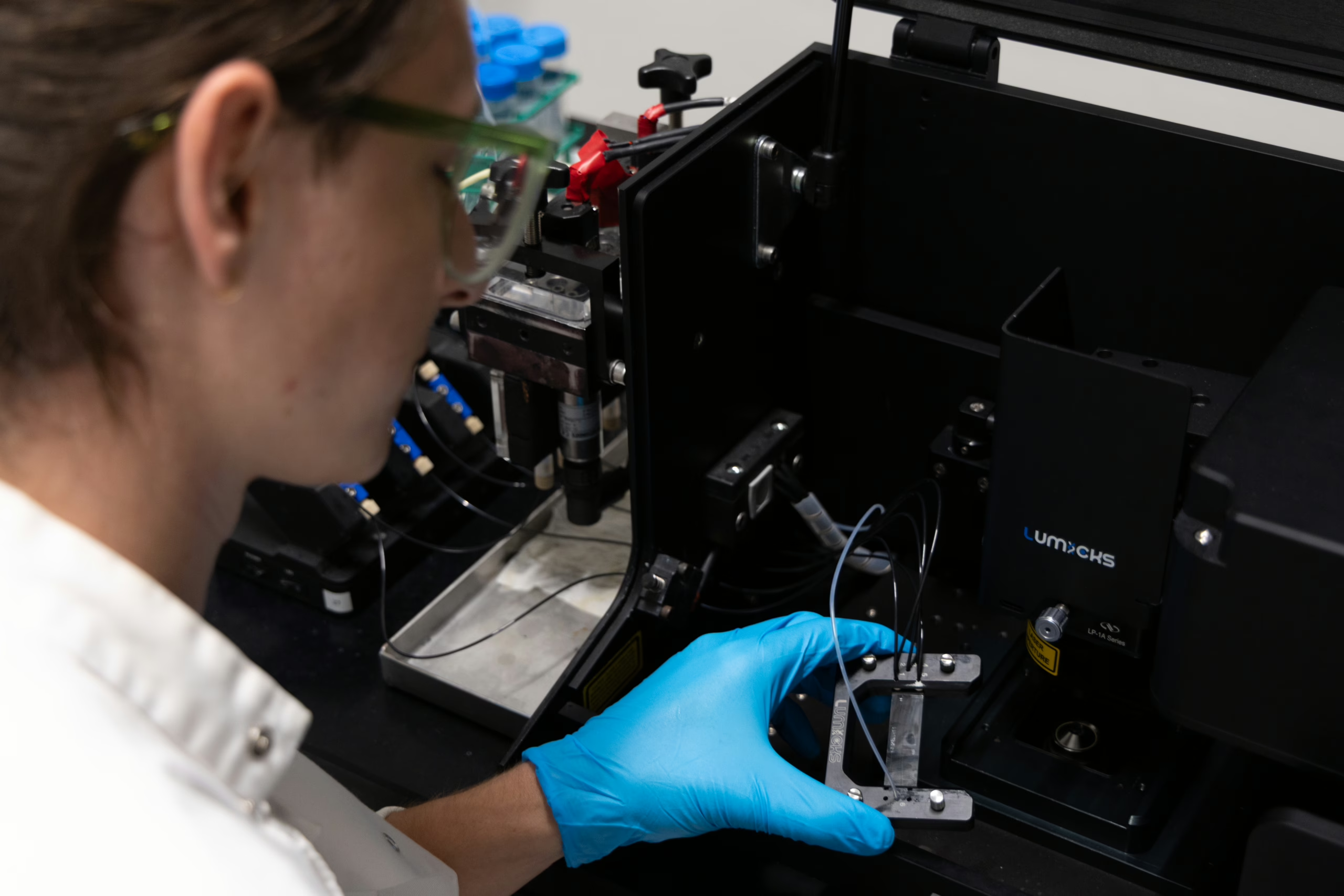
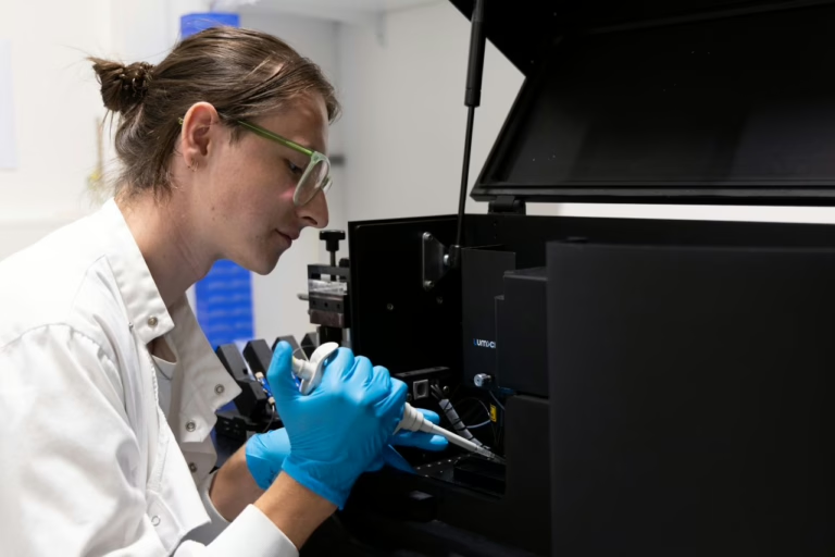
At the molecular level, we use optical tweezers and single-molecule fluorescence to study how chaperones fold amino-acid chains into functional proteins, and to probe ribosomes and translation dynamics.
At the cellular level, we use 3D time-lapse microscopy and AI-driven image analysis to understand how multi-cellular systems self-organize, with a specific interest in organoids.












Biophysics Latest news

Most recent publications